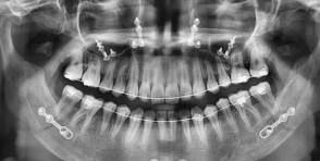

Teeth imaging is an important part of modern dental care. Dentists use different imaging methods to see areas of the teeth, jaw, and surrounding structures that cannot always be examined during a regular dental checkup. These images can help dental professionals understand what is happening below the surface and make better decisions about treatment.
A routine dental examination allows a dentist to inspect visible areas of the mouth. However, some problems develop between teeth, underneath existing fillings, inside the jawbone, or around the roots. Teeth imaging can provide additional information when a visual examination alone is not enough.
Dental imaging may be recommended during a routine examination, before certain procedures, or when a patient has symptoms such as tooth pain, swelling, sensitivity, or an injury. The type of image used depends on the patient’s situation and what the dentist needs to examine.
What Is Teeth Imaging?
Teeth imaging refers to techniques used to create pictures of the teeth, jawbone, and other oral structures. The most familiar form is the dental X-ray, but modern dentistry also uses more advanced imaging systems.
Traditional dental X-rays use a small amount of radiation to create images of the teeth and surrounding structures. Other systems can produce wider or three-dimensional views, allowing dentists to examine areas from different angles.
The main purpose is to provide information that may not be visible during a standard dental examination. For example, an image may show a cavity between two teeth, changes around a tooth root, an impacted tooth, or changes in the supporting bone.
The dentist can then combine the imaging results with the patient’s symptoms, dental history, and physical examination.
Why Do Dentists Use Teeth Imaging?
Dentists use imaging for many different reasons. One common purpose is to look for dental problems that cannot be seen easily during a visual examination.
Imaging can help identify cavities between teeth, infections around roots, bone changes, impacted teeth, and other conditions. It may also help a dentist plan procedures such as tooth extraction, dental implants, root canal treatment, or orthodontic care.
Children and teenagers may receive imaging when their dentist needs to monitor developing teeth. Adults may need images when there are symptoms or when a dentist is planning a particular treatment.
Not every patient needs the same type or frequency of imaging. A dental professional usually considers the patient’s age, symptoms, medical and dental history, previous images, and current treatment needs before recommending an examination.
Common Types of Dental X-Rays
Several types of dental X-rays are commonly used. Each one provides a different view of the mouth.
Bitewing X-rays show the crowns of the upper and lower teeth in a particular area. They are especially useful for detecting cavities between teeth and evaluating the level of supporting bone.
Periapical X-rays show an entire tooth, including the crown, root, and surrounding bone. Dentists may use them when investigating pain, infection, or changes around a tooth’s root.
Panoramic X-rays provide a broad view of the upper and lower jaws, teeth, and surrounding structures. They can be useful for assessing impacted wisdom teeth, orthodontic concerns, jaw problems, and overall dental development.
Occlusal X-rays can show a larger area of the mouth and may be used when a dentist needs a broader view of developing or impacted teeth.
The choice depends on what the dentist needs to evaluate.
Bitewing Imaging
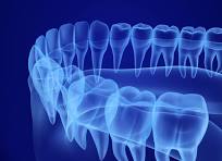
Bitewing imaging is one of the common methods used during dental examinations. The patient typically bites gently on a small holder while the X-ray image is taken.
The resulting image can show the crowns of several upper and lower teeth at once. It is particularly useful for finding cavities that may develop in areas where teeth touch each other.
These areas can be difficult to inspect directly, even when a dentist performs a careful examination.
Bitewing images may also help dentists evaluate the level of bone supporting the teeth. Changes in bone levels can provide useful information when assessing periodontal health.
Periapical Imaging
A periapical X-ray provides a detailed view of an individual tooth or a small group of teeth. It includes the visible crown as well as the root and surrounding bone.
Dentists may request this type of imaging when a patient reports persistent tooth pain or when an infection is suspected.
The image can help show changes around the root that may not be visible inside the mouth. It can also assist with treatment planning for procedures involving the tooth root.
Because it provides a focused view, periapical imaging can be useful when a dentist needs detailed information about a specific area.
Panoramic Dental Imaging
A panoramic X-ray provides a broad image of the mouth and jaw. Unlike a small intraoral X-ray, it captures a larger area in one image.
This type of imaging can be useful when dentists need an overall view of the teeth and jaw structures. It may be used when evaluating wisdom teeth, orthodontic development, jaw conditions, or certain dental abnormalities.
Panoramic imaging can also help dentists plan some procedures by providing a general overview of the structures involved.
However, a panoramic image does not replace every other type of dental X-ray. If a dentist needs a highly detailed view of one tooth, another imaging method may be more appropriate.
CBCT Dental Imaging
Cone beam computed tomography, commonly called CBCT, is an advanced imaging technique that can produce three-dimensional views of dental and facial structures.
A CBCT scan can provide information from multiple angles. This can be particularly useful when a dentist or oral health specialist needs detailed information for treatment planning.
CBCT may be considered for certain dental implant procedures, complex tooth extractions, orthodontic planning, root canal assessment, or evaluation of particular jaw structures.
Because CBCT uses radiation and provides more information than a standard dental X-ray, it is generally used when the additional detail is considered useful for the patient’s care.
How Teeth Imaging Helps Detect Cavities
Cavities do not always appear clearly during a visual dental examination. Some develop in areas between teeth where a dentist cannot easily see the affected surface.
Dental imaging can help reveal these hidden areas.
Bitewing X-rays are commonly used to identify cavities between teeth. Finding a problem early may allow a dentist to recommend treatment before the damage becomes more extensive.
However, an X-ray is only one part of the diagnostic process. Dentists also consider visual findings, symptoms, previous dental history, and other information when evaluating possible tooth decay.
Teeth Imaging for Root Problems
Problems involving tooth roots can sometimes be difficult to identify through a visual examination alone.
A periapical X-ray can show the root and surrounding bone. This can help a dentist investigate symptoms such as persistent pain, swelling, or sensitivity.
Imaging may also be used during root canal treatment planning. The dentist can examine the shape and position of the root and look for changes around the surrounding bone.
The information from the image is then combined with clinical findings to determine the appropriate treatment.
Imaging for Impacted Teeth
Some teeth do not emerge normally through the gums. Wisdom teeth are a common example.
A panoramic X-ray can help dentists see the position of developing or impacted teeth. This information can be useful when deciding whether monitoring, removal, or another form of treatment may be appropriate.
Three-dimensional imaging may be considered in more complex cases where the dentist needs a detailed understanding of the relationship between a tooth and nearby structures.
The exact imaging method depends on the patient’s individual situation.
Teeth Imaging for Dental Implants
Dental implants require careful planning because the implant must be positioned in an appropriate location within the jaw.
Imaging can help the dental team evaluate the available bone and understand the position of nearby anatomical structures.
In some cases, three-dimensional imaging such as CBCT can provide information that a standard X-ray cannot provide.
The dentist can use this information to plan the procedure and determine the appropriate approach.
Not every implant case requires the same imaging. The dental professional decides which method provides enough information for safe and effective planning.
Teeth Imaging During Orthodontic Treatment
Orthodontists may use dental imaging to understand the position of teeth and the development of the jaw.
X-rays can show teeth that have not erupted yet, the position of impacted teeth, and certain changes in the supporting structures.
This information can help an orthodontist develop an appropriate treatment plan.
Imaging may also be used during treatment when there is a specific reason to check progress or investigate an unexpected problem.
The goal is to obtain useful information while avoiding unnecessary exposure.
What Happens During a Dental X-Ray?
A standard dental X-ray is usually a quick process.
The patient may be asked to sit or stand while the dental professional positions the imaging equipment. For an intraoral X-ray, a small sensor or film is placed inside the mouth.
The patient may need to bite gently to keep the sensor in position.
The equipment is then positioned outside the mouth, and the image is captured. The process normally takes only a short amount of time.
Several images may be taken if the dentist needs to examine different areas.
Most dental X-rays do not require special preparation.
What Happens During a Panoramic Scan?
A panoramic scan is also relatively straightforward.
The patient generally stands or sits in a specific position and places their chin or head against a support. The dental professional explains how to remain still during the scan.
The imaging equipment moves around the head while capturing the required image.
The process is generally quick, and the patient does not need to stay still for a long period.
The resulting image gives the dental professional a broad view of the teeth and jaws.
Is Teeth Imaging Safe?
Dental X-rays use radiation, so dentists generally follow the principle of obtaining an image when it is clinically useful.
Modern dental imaging equipment is designed to use relatively low levels of radiation. Dental professionals also use appropriate techniques and protective measures to reduce unnecessary exposure.
The benefits of obtaining useful diagnostic information are considered alongside the small radiation exposure involved.
Patients who are pregnant or may be pregnant should inform their dental professional before imaging. The dentist can then consider the situation and determine the appropriate approach.
Patients should also tell their dentist about previous dental images when relevant because avoiding unnecessary duplicate imaging can reduce exposure
Digital imaging has become common in modern dental practices. With digital X-ray systems, dental professionals can produce and view images electronically within a short time. These images can also be stored securely and compared with future scans when appropriate. This technology makes it easier for dental teams to review and manage a patient’s imaging history.
Another advantage is that digital systems can make it easier for dental professionals to share images with specialists when a referral is needed.
The technology used by each dental practice may vary, but digital imaging has become an important part of modern dental care.
Teeth Imaging and Dental Emergencies
Dental imaging can be especially useful when a patient has experienced an injury.
A damaged tooth may have a crack or root problem that cannot be fully evaluated through a visual examination.
An X-ray can provide additional information about the tooth and surrounding structures.
For example, imaging may help a dentist assess an injured tooth, evaluate surrounding bone, or look for other problems caused by trauma.
In an emergency, the dentist decides whether imaging is necessary based on the injury and symptoms.
Dental Imaging for Children
Children may need dental imaging for several reasons.
Developing permanent teeth are not always visible in the mouth, so imaging can help dentists monitor dental development.
X-rays may also help identify cavities or other issues that are difficult to see during a routine examination.
Because children are more sensitive to radiation than adults, dental professionals generally use imaging carefully and only when there is a clinical reason.
Parents can ask the dental team why a particular image is recommended and what information it will provide.
How Often Should Dental X-Rays Be Taken?
There is no single schedule that applies to every patient.
The need for dental imaging depends on factors such as age, oral health, symptoms, risk of dental disease, previous findings, and treatment needs.
Someone with a history of dental problems may require imaging at different intervals from someone with a low risk of disease.
Dentists consider the patient’s individual circumstances rather than automatically taking the same images at every visit.
This approach helps ensure that imaging provides useful information without unnecessary examinations.
Preparing for Teeth Imaging
Most routine dental X-rays require little or no preparation.
Patients may simply need to remove glasses, jewelry, or other objects that could interfere with the image.
The dental professional will explain what to do during the examination.
For certain advanced imaging procedures, additional instructions may be provided.
Patients should also mention relevant medical information and tell the dental team if they are pregnant or might be pregnant.
Following the instructions carefully can help the imaging process run smoothly.
Understanding Your Dental Images
Dental X-rays can look complicated to someone who has never studied them.
Teeth appear as lighter structures, while areas with less density appear darker.
A dentist has the training needed to interpret these images in combination with a clinical examination.
Patients should avoid trying to diagnose themselves based only on an image found online or a copy of their own dental X-ray.
If a dentist identifies an area of concern, they can explain what the image shows and how it relates to the patient’s symptoms.
Benefits of Modern Teeth Imaging
Modern dental imaging provides several benefits.
It can help dentists detect problems earlier, understand conditions that are not visible during a regular examination, and plan treatments more accurately.
Three-dimensional imaging can provide additional information for selected complex procedures.
Digital systems can also make it easier to store, compare, and share images when appropriate.
These benefits can improve the overall diagnostic process and help dental professionals provide more informed care.
Limitations of Dental Imaging
Dental imaging is useful, but it does not show everything.
Different imaging techniques provide different types of information. A standard X-ray may be excellent for certain dental problems but may not provide the detailed three-dimensional information needed for a complex procedure.
Images must also be interpreted alongside a clinical examination.
A dentist may need additional tests or another type of imaging when the first image does not provide enough information.
Therefore, dental imaging should be viewed as one part of comprehensive dental care rather than a complete diagnosis by itself.
How Technology Is Improving Dental Imaging
Technology continues to improve dental imaging.
Modern systems can produce clearer images while supporting efficient workflows. Three-dimensional imaging has also expanded the information available for selected dental procedures.
Computer software can help dental professionals view images from different angles and examine specific areas more closely.
Digital records also make it easier to maintain a patient’s imaging history.
As technology continues to develop, dental professionals may have access to increasingly useful tools for diagnosis and treatment planning.
Choosing the Right Dental Imaging Method
The best imaging method depends on the clinical question.
If a dentist wants to check for cavities between teeth, bitewing imaging may be appropriate.
the concern involves the root of one tooth, a periapical image may provide better information.
If a broad view of the jaws is needed, panoramic imaging may be useful.
For certain complex cases, a CBCT scan may provide the three-dimensional information required for treatment planning.
The dentist chooses the method based on the patient’s needs rather than simply selecting the most advanced technology available.
Questions to Ask Your Dentist
Patients can ask questions about any recommended dental imaging procedure.
Useful questions include:
- Why do I need this image?
- What problem are you checking for?
- What type of imaging will be used?
- How will the results affect my treatment?
- Do you need to compare this image with an older one?
- Are there alternative ways to obtain the information?
Understanding the reason for an examination can help patients feel more comfortable and involved in their dental care.
Maintaining Good Oral Health
Teeth imaging is useful for detecting and evaluating dental problems, but prevention remains important.
Regular brushing and flossing help remove plaque and reduce the risk of tooth decay and gum disease.
A balanced diet and sensible choices around sugary foods and drinks can also support oral health.
Regular dental examinations allow professionals to identify potential problems and provide advice based on individual needs.
When imaging is necessary, it adds another source of information that can help the dental team understand the condition of the teeth and surrounding structures.
Final Thoughts
Teeth imaging has become an important part of modern dental care. From simple bitewing X-rays to panoramic scans and advanced three-dimensional imaging, different techniques provide dentists with valuable information about teeth, roots, jaws, and surrounding structures.
Dental imaging can help identify problems that may not be visible during a normal examination. It can also support treatment planning for procedures such as root canal treatment, orthodontics, tooth removal, and dental implants.
The right imaging method depends on the patient’s individual needs. Dentists consider symptoms, dental history, age, existing conditions, and the information required before recommending an examination.
If your dentist recommends dental imaging, asking why the image is needed and how the results will affect your care can help you understand the process. With appropriate use, modern dental imaging can provide useful information and support better-informed dental care.